原子力显微镜
AFM配件
应用
联系我们
 牛津仪器集团成员

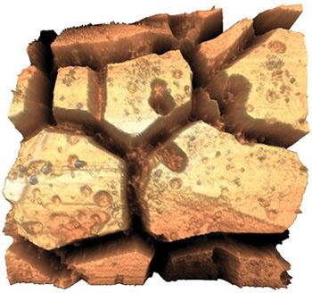
利用原子力显微镜,科研人员能够对生物材料的形貌和机械特性进行表征。可以准确地测量材料表面的粗糙度和微观结构随着成分和工艺的变化。在体外试验或者移植后,同样可以对其进行观测,以评估表面特征的变化。此外,科研人员还可以利用多种技术来测量生物材料的刚度、模量和耗散。
咨询AFM领域的专家"Silk protein nanowires patterned using electron beam lithography," R. K. Pal, and V. K. Yadavalli, 29, 335301 (2018). https://doi.org/10.1088/1361-6528/aac855
"Type I collagen from jellyfish Catostylus mosaicus for biomaterial applications," Z. Rastian, S. Pütz, Y. J. Wang, S. Kumar, F. Fleissner, T. Weidner, and S. H. Parekh, ACS Biomater. Sci. Eng. 4, 2115 (2018). https://doi.org/10.1021/acsbiomaterials.7b00979
"Mutable polyelectrolyte tube arrays: mesoscale modeling and lateral force microscopy," S. W. Cranford, L. Han, C. Ortiz, and M. J. Buehler, Soft 13, 5543 (2017). https://doi.org/10.1039/c7sm00864c
"Engineered phage nanofibers induce angiogenesis," S. Y. Yoo, K. R. Shrestha, S. N. Jeong, J. I. Kang, and S. W. Lee, Nanoscale 9, 17109 (2017). https://doi.org/10.1039/c7nr03332j
"3D bioprinted oxide-incorporated matrix for promoting chondrogenic differentiation of human bone marrow mesenchymal stem cells," X. Zhou, M. Nowicki, H. Cui, W. Zhu, X. Fang, S. Miao, S. J. Lee, M. Keidar, and L. G. Zhang, Carbon 116, 615 (2017). https://doi.org/10.1016/j.carbon.2017.02.049
"The structure and mechanical properties of articular cartilage are highly resilient towards transient dehydration," K. Boettcher, S. Kienle, J. Nachtsheim, R. Burgkart, T. Hugel, and O. Lieleg, Acta Biomater. 29, 180 (2016). https://doi.org/10.1016/j.actbio.2015.09.034
"Heterogeneous silicon mesostructures for lipid-supported bioelectric interfaces," Y. Jiang, J. L. Carvalho-de-Souza, R. C. Wong, Z. Luo, D. Isheim, X. Zuo, A. W. Nicholls, I. W. Jung, J. Yue, D. J. Liu, and Y. Wang, Nat. Mater. 15, 1023 (2016). https://doi.org/10.1038/nmat4673
"Regenerated silk materials for functionalized silk orthopedic devices by mimicking natural processing," C. Li, B. Hotz, S. Ling, J. Guo, D. S. Haas, B. Marelli, F. Omenetto, S. J. Lin, and D. L. Kaplan, Biomaterials 110, 24 (2016). https://doi.org/10.1016/j.biomaterials.2016.09.014
"Modulation of protein fouling and interfacial properties at carbon surfaces via immobilization of glycans using aryldiazonium chemistry," F. Zen, M. D. Angione, J. A. Behan, R. J. Cullen, T. Duff, J. M. Vasconcelos, E. M. Scanlan, and P. E. Colavita, Sci. Rep. 6, 24840 (2016). https://doi.org/10.1038/srep24840
"Nanomechanics of cells and biomaterials studied by atomic force microscopy," J. I. Kilpatrick, I. Revenko, and B. J. Rodriguez, Adv. Healthcare Mater. 4, 2456 (2015). https://doi.org/10.1002/adhm.201500229
"The preparation and characterization of polycaprolactone/ oxide biocomposite nanofiber scaffolds and their application for directing cell behaviors," J. Song, H. Gao, G. Zhu, X. Cao, X. Shi, and Y. Wang, Carbon 95, 1039 (2015). https://doi.org/10.1016/j.carbon.2015.09.011
"Application of layer-by-layer coatings to tissue scaffolds—development of an angiogenic biomaterial," C. D. Easton, A. J. Bullock, G. Gigliobianco, S. L. McArthur, and S. MacNeil, J. Mater. Chem. B 2, 5558 (2014). https://doi.org/10.1039/c4tb00448e
"Chirality effects at each amino acid position on tripeptide self-assembly into hydrogel biomaterials," S. Marchesan, C. D. Easton, K. E. Styan, L. J. Waddington, F. Kushkaki, L. Goodall, K. M. McLean, J. S. Forsythe, and P. G. Hartley, Nanoscale 6, 5172 (2014). https://doi.org/10.1039/c3nr06752a
"Load-bearing in cortical bone microstructure: Selective stiffening and heterogeneous strain distribution at the lamellar level," O. L. Katsamenis, H. M. Chong, O. G. Andriotis, and P. J. Thurner, J. Mech. Behav. Biomed. Mater. 17, 152 (2013). https://doi.org/10.1016/j.jmbbm.2012.08.016
"Nanomechanical mapping of the osteochondral with contact resonance force microscopy and nanoindentation," S. E. Campbell, V. L. Ferguson, and D. C. Hurley, Acta Biomater. 8, 4389 (2012). https://doi.org/10.1016/j.actbio.2012.07.042
"Dilatational band formation in bone," A. A. Poundarik, T. Diab, G. E. Sroga, A. Ural, A. L. Boskey, C. M. Gundberg, and D. Vashishth, Proc. Natl. Acad. Sci. U.S.A. 109, 19178 (2012). https://doi.org/10.1073/.1201513109
"Immunofluorescence-guided atomic force microscopy to measure the micromechanical properties of the pericellular matrix of porcine articular cartilage," R. E. Wilusz, L. E. DeFrate, and F. Guilak, J. Royal Soc. 9, 2997 (2012). https://doi.org/10.1098/rsif.2012.0314
"Diameter of titanium nanotubes influences anti-bacterial efficacy," B. Ercan, E. Taylor, E. Alpaslan, and T. J. Webster, 22, 295102 (2011). https://doi.org/10.1088/0957-4484/22/29/295102
"Experimental evidence for interfacial biochemical bonding in osseointegrated titanium implants," Y.-T. Sul, D. H. Kwon, B.-S. Kang, S.-J. Oh, and C. Johansson, Clin. Oral Implants Res. 24, 8 (2011). https://doi.org/10.1111/j.1600-0501.2011.02355.x
"Physical and electromechanical properties of doped polypyrrole biomaterials," A. Gelmi, M. J. Higgins, and G. G. Wallace, Biomaterials 31, 1974 (2010). https://doi.org/10.1016/j.biomaterials.2009.11.040
"Autonomic self‐healing of hydrogel films," A. B. South and L. A. Lyon, Angew. Chem. Int. Ed. 49, 767 (2010). https://doi.org/10.1002/anie.200906040
"Titanium implant modification by cathodic reduction in hydrofluoric acid: characterization and in vivo performance," S. F. Lamolle, M. Monjo, S. P. Lyngstadaas, J. E. Ellingsen, and H J. Haugen, J. Biomed. Mater. Res. 88A, 581 (2009). https://doi.org/10.1002/jbm.a.31898
"Hydride formation on titanium surfaces by cathodic polarization," K. Videm, S. Lamolle, M. Monjo, J. E. Ellingsen, S. P. Lyngstadaas, and H J. Haugen, Appl. Surf. Sci. 255, 3011 (2008). https://doi.org/10.1016/j.apsusc.2008.08.090
"Protective coatings on extensible biofibres," N. Holten-Andersen, G. E. Fantner, S. Hohlbauch, J. H. Waite, and F. W. Zok, Nat. Mater. 6, 669 (2007). https://doi.org/10.1038/nmat1956
"Nanoscale heterogeneity promotes energy dissipation in bone," K. Tai, M. Dao, S. Suresh, A. Palazoglu, and C. Ortiz, Nat. Mater. 6, 454 (2007). https://doi.org/10.1038/nmat1911

© 牛津仪器 2026
 公安机关备案号31010402003473